Mangrove-associated Algae from Samoa, South Pacific

Posa A. Skelton and G. Robin South
FIGURES

plate 1

Map of Samoa


plate 1

Plate I. Figs 1-7: Cyanophytes.
Fig. 1: Lyngbya sp. 1, scale = 27µm. Fig. 2: Lyngbya sp. 3, scale = 25µm. Fig. 3: Lyngbya sp. 2, scale = 50µm. Fig. 4: Calothrix confervicola habit, scale = 100µm. Fig. 5: C. confervicola, trichomes showing numerous intercalary heterocytes, scale = 25µm. Fig. 6: Schizothrix cf. tenerrima, scale = 12µm. Fig. 7. Calothrix crustacea, showing false (Scytonema-like) branching, scale = 12.5 µm


plate 2

Plate II. Figs 8-13: Ceramium spp. Figs. 8, 10, 12: Ceramium flaccidum. Fig. 8: habit, scale = 50µm. Fig. 10: nodes and internodes with elongated basipetal cortical cells (arrows), distinctive of this species, scale = 15µm. Fig. 12: unicellular rhizoid cut off from pericentral cell, scale = 50µm. Figs. 9, 11, 13: Ceramium upolense. Fig. 9: habit, scale = 200µm. Fig. 11: nodal cortication, scale = 50µm. Fig. 13: two or more rhizoids cut off from periaxials, scale = 200µm.


plate 3

Plate III.
Figs. 14-19: Caloglossa adhaerens. Fig. 14: habit, scale = 2mm. Fig. 15: branch tips, scale = 500µm. Figs 16-17: antheridial branch, showing sterile marginal cells, scales = 200µm & 150µm, respectively. Fig. 18: tetrasporic branch, scale = 1mm. Fig. 19: rhizoids arising from second and third order cells terminating in haptera or blunt tips, scale = 100µm.


plate 4

Plate IV. Figs. 20-28: Caloglossa leprieurii.
Fig. 20: habit showing various plants, scale = 1cm. Fig. 21: cross section of stipe, scale = 200µm. Fig. 22: habit of an individual plant, scales = 2mm. Fig. 23: branches arising from the constricted node often issuing a basal rhizoid, scale = 0.5mm. Fig. 24: cross section of blade with four pericentral cells, scale = 150µm. Fig. 25: heavily corticated rhizoid forming a short stipe, scale = 1.5mm. Fig. 26: tetrasporangial sorus, scale = 0.5mm. Fig. 27: a cystocarp arising near the apex of a blade, scale = 0.5mm. Fig. 28: spermatangial sorus on both sides of the blade (dark patches), scale = 0.5mm.


plate 5

Plate V. Figs. 29-33: Enteromorpha clathrata.
Fig. 29: habit with numerous branchlets, scale = 1.5mm. Fig. 30: cell arrangements of a young branch, scale = 1mm. Fig. 31: cell arrangement of mature branch, scale = 500µm. Fig. 32: tips of branches, scale = 1mm. Fig. 33: multicellular rhizoids, scale = 1mm.


plate 6

Plate VI. Figs. 34-43.
Fig. 34: Enteromorpha clathrata, young plant showing uniseriate (arrow) and multiseriate branches, scale = 50µm;
Figs. 35-36: Chaetomorpha brachygona, rectangular shaped cells, scale = 150µm;
Figs. 37-41: Chaetomorpha minima. Fig. 37: habit, scale 250µm. Fig. 38: basal cell, scale = 50µm. Fig. 39: close up of long attenuating basal cell (arrow), scale = 50µm. Fig. 40: slightly swollen tip, scale 25µm. Fig. 41: filament cells of various shape, scale = 25µm;
Figs. 42-43: Cladophora liebtruthii. Fig. 42: habit, scale = 500µm. Fig. 43: digitate holdfast attached to another filament, scale = 100µm.


plate 7

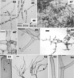
Plate VII. Figs. 44-54.
Figs. 44-46: Rhizoclonium africanum Figs. 44-45: filaments producing short rhizoids (arrows), scales = 100µm. Fig. 46 fine filaments, scale = 100µm.
Figs. 47-51: Boodlea composita. Fig. 47: habit, scale = 1mm. Fig. 48: branching, scale = 400µm. Fig. 49: branching showing rhizoidal holdfast (arrow), scale = 50µm. Fig. 50: branching pattern, scale = 250µm. Fig. 51: showing crystals (arrows), scale = 250µm.
Figs. 52-53: Cladophoropsis carolinensis. Fig. 52: habit of two plants, scale = 1mm; Fig. 53: tip, scale = 100µm. Fig. 54: branching with opening, scale = 100µm.