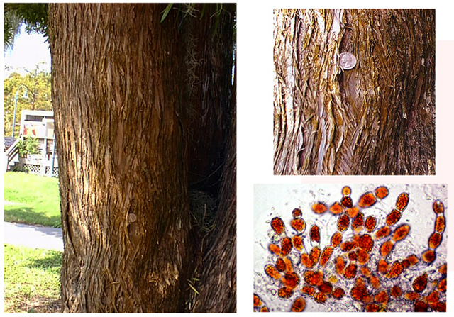
Fig. 1. Habit of Trentepohlia sp. growing as a corticolous form in Louisiana.

Fig. 2. Magnolia leaves from Louisiana. Black spots associated with Cephaleuros parasiticus, a "parasitic" species of Trentepohliaceae.

Fig. 3. Trentepohlia sp. Isolated from bark of Magnolia tree growing on University of Louisiana at Lafayette campus.

Fig. 4. Discoid thallus in Phycopeltis sp.

Fig. 5. Branched filaments of Trentepohlia sp. Sample from Louisiana, USA, rich in burnt orange droplets containing carotenoids.

Fig. 6. Sporangia of Cephaleuros, SP=sporangiophore; HC=head cell; SC= suffultory cell; S=sporangium. The suffultory cell and sporangium together form the sporangiate-lateral (SL).

Fig. 7. Sporangiate lateral in Cephaleuros virescens consisting of a sporangium and a suffultory cell. Plasmodesmata can be seen at the cross walls of the point of attachment (double ring).

Fig. 8. Magnolia leaves from Louisiana with Cephaleuros spp. infestation. Lichenized Cephaleuros thalli are evident as white spots on the leaves.

Fig. 9. Habitat and habit of a corticolous Trentepohlia sp. on Magnolia tree from the University of Louisiana at Lafayette campus, USA.

Fig. 10. Printzina lagenifera. Filaments from liquid culture. (Photo F. Rindi and M. Guiry)

Fig. 11. Phycopeltis arundinacea. Thallus with a discoid shape. (Photo F. Randi and M. Guiry).
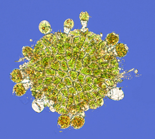
Fig. 12. Phycopeltis epiphyton. Small discoid thallus showing marginal sporangiate laterals. (Photo F. Rindi and M. Guiry.)

Fig. 13. Cross section of Magnolia leaf showing subcuticular thallus of Cephaleuros virescens with protruding reproductive structures. The conspicuous head with sporangia is subtended by a long and narrow "tail", or sporangiophore.

Fig. 14. Cross section of a Magnolia leaf showing a subcuticular thallus of Cephaleuros virescens.

Fig. 15. Cephaleuros virescens, sporangiophore with inflated head subtending the sporangiate laterals.
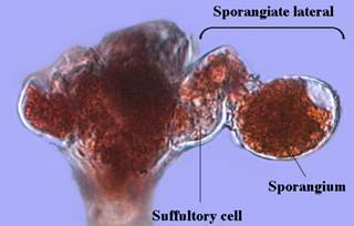
Fig. 16. Cephaleuros virescens. Sporangiate lateral showing a suffultory cell and a sporangium; a papilla (exit pore) can be seen near the attachment area of the sporangium.

Fig. 17. Stomatochroon sp. Several sporangiate laterals are evident on the head cell.

Fig. 18. Stomatochroon sp. Sporangiate lateral with an extended suffultory cell and a sporangium.

Fig. 19. Sporangium of Stomatochroon sp. Note papilla pore close to attachment area.

Fig. 20. Cultures of specimens of Trentepohliales. The isolates are maintained with constant fluorescent illumination at room temperature (ca. 20 C). Culture media are solid (agarized) or liquid.

Fig. 21. Location of primers used in PCR amplification of the nuclear 18SSU rDNA.
